Multiscale Structural Dynamics of p53 Intrinsically Disordered Regions (IDRs)
Project Overview
This research utilized extensive molecular dynamics (MD) simulations to explore the conformational landscapes of all four intrinsically disordered regions (IDRs) within the human tumor suppressor protein p53: TAD1 (transactivation domain 1), PRD (proline-rich domain), PTL (pre-tetramerization loop), and REG (regulatory domain). The study revealed how local sequence features, post-translational modifications (PTMs), and physical constraints imposed by adjacent globular domains dictate the dynamic behavior essential for p53’s function.
I. TAD1: Transactivation Domain 1 (M1–E28)
The Transactivation Domain 1 (TAD1), an intrinsically disordered region (IDR) spanning residues 1-40 in p53, is the primary functional segment responsible for interacting with basal transcription machinery and key regulatory proteins. Its high flexibility and conformational plasticity are essential for its functional versatility. Critically, TAD1’s N-terminus is the target of MDM2, the critical E3 ubiquitin ligase and primary negative regulator of p53, which targets p53 for degradation.
The p53-MDM2 Binding Interface
The high affinity interaction between p53 and MDM2 is mediated by a conserved 12-residue motif (L14–L26) on TAD1.
- Induced Conformational Change: In its free state, TAD1 is intrinsically disordered. However, upon binding to MDM2, the 12-residue motif must undergo an induced fit, transiently adopting an $\alpha$-helical conformation (particularly residues T18–L25) that docks into a hydrophobic pocket within MDM2’s SWIB domain.
- Key Contact Residues: This stabilization is driven by three essential hydrophobic residues on the p53 peptide: F19, W23, and L26. Mutations in any of these hydrophobic residues can directly impair binding and alter p53 regulation.
The Molecular Switch: Phosphorylation at S15 and T18
Phosphorylation at Ser15 (S15) and Thr18 (T18) is known to inhibit p53-MDM2 binding. Molecular dynamics (MD) studies of the isolated TAD1 domain modeled how double phosphorylation acts as a molecular switch to achieve this inhibition.
Conformational Dynamics in Unbound TAD1
MD studies modeled three states of the M1-E28 segment of TAD1 using the validated AMBER99SB-DISP (DISP) force field: APO (non-phosphorylated), pS15 (singly phosphorylated), and pS15/pT18 (doubly phosphorylated).
- pS15 State: Single phosphorylation at pS15 had minimal impact on the overall shape of the protein and left the contact map largely unchanged compared to the APO state.
- pS15/pT18 State: Double phosphorylation forced the domain into a significantly more compact structure. This compaction was exhibited by a strong bifurcation in both the RMSD and $R_g$ profiles.
Inhibition Mechanism: From Helix to Sheet
The compaction caused by the pS15/pT18 state actively prevents the required $\alpha$-helix formation and therefore inhibits MDM2 binding.
- Disruption of $\alpha$ Helices: Double phosphorylation diminishes the $\alpha$-helices in the region near the phosphorylation sites.
- Stabilization of $\beta$ Sheets: This highly condensed state facilitates long-range interactions, stabilizing a long-range $\beta$-sheet structure between residues V10 and W23. This competing structure physically blocks the key hydrophobic residues necessary to form the binding $\alpha$-helix.
- Reduced Binding Affinity: The functional consequence is a reduced binding affinity ($\Delta G_{binding}$) in the double phosphorylated complex. Calculations showed a decrease from an average $\Delta G_{binding}$ of $-36.4\ \text{kcal}/\text{mol}$ (APO) to $-22.9\ \text{kcal}/\text{mol}$ (phosphorylated).
Methodology Insights
To accurately model this IDR, four force fields were benchmarked against experimental NMR chemical shift data.
- Optimal Force Field Selection: The AMBER99SB-DISP (DISP) force field was identified as the optimal and most versatile choice, showing the best overall agreement with experimental NMR chemical shift values (highest $r^2$ values and low RMSE across atom types).
- Dynamic Residue Network Analysis (DRNA): A Dynamic Residue Network Analysis (DRNA) approach was introduced to understand how the phosphorylation signal propagates through the complex. This technique maps allosteric communication pathways by quantifying the correlated motions (edges) between residues (nodes). This provided mechanistic insight into the allosteric pathways disrupted by the phosphorylation switch.
II. PRD: Proline-Rich Domain (Residues 64–92)
The PRD acts as a critical linker domain, characterized by high proline content and PXXP motifs involved in SH3 domain binding and apoptotic signaling.
- Wild-Type Structure: The unbound PRD ensemble is predominantly composed of unordered structure (45.1%) and transient polyproline II (PPII) helices (31.1%). The high proline content imposes local rigidity, restricting backbone motion.
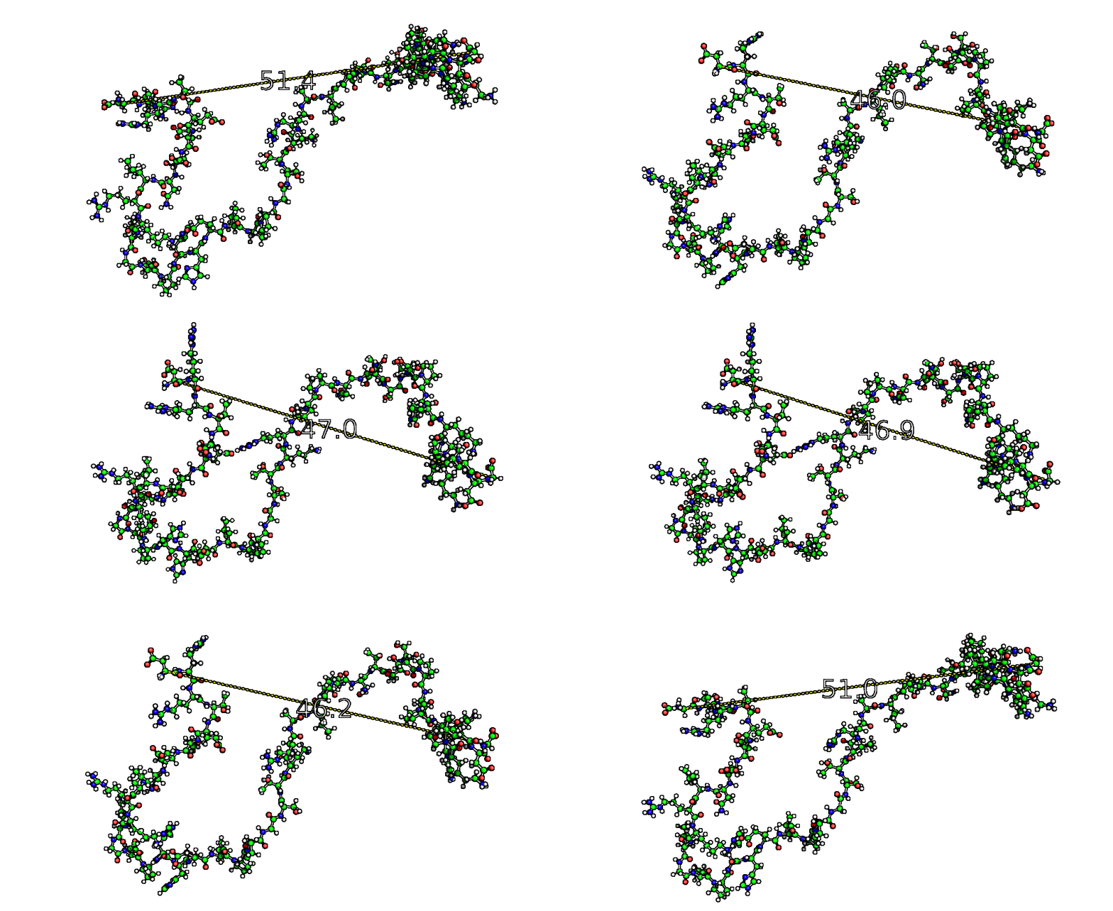
- Structural Robustness (IDR vs. IDP): Restraining the termini to mimic the physiological context (IDR model) greatly improved agreement with experimental SAXS data ($\chi^{2}=0.056$ at $\mathbf{R_{ee}} = 51.2\ \text{Å}$). Crucially, the domain’s fundamental PPII secondary structure profile remained largely unaffected by the physical constraint.
- Mutational Impact:
- P72R: This common polymorphism disrupts a consecutive proline pair (P71-P72), increasing local conformational flexibility.
- P82L: This mutation abolishes a conserved PXXP motif, eliminating the associated PPII helix and interfering with the PIN1-dependent cis-trans isomerisation required for Ser20 phosphorylation and p53 activation.
III. PTL: Pre-Tetramerization Loop (Residues 281–325)
The Pre-Tetramerization Loop (PTL), spanning residues $\mathbf{Lys_{292}}$ to $\mathbf{Gly_{325}}$, is an intrinsically disordered region (IDR) crucial for the human tumor suppressor protein p53’s oligomerization process. Its inherent flexibility provides the necessary conformational changes for tetramerization and significantly increases DNA binding affinity (1000-fold).
Molecular Dynamics (MD) Investigation Approach
To explore the PTL’s conformational dynamics, we performed atomistic MD simulations using the GROMACS package and the AMBERSB99-ILDN force field with the TIP4P-D water model, which is well-suited for simulating IDRs.


- Unrestrained Trajectory (PTL as an IDP):
- Simulated as an independent, free-moving intrinsically disordered protein (IDP).
- The Radius of Gyration ($\mathbf{R_g}$) comfortably settled around $\mathbf{1.8\ \text{nm}}$ but could naturally expand up to $3.4\ \text{nm}$ in its unrestrained form.
- The dimensionless Kratky plot indicated a highly flexible and disordered region, sampling a wide range of phase space.
- The predominant secondary structures sampled were $\mathbf{Polyproline\ Type\ II\ (PPII)\ helices}$ ($\sim 19\%$ detected by DSSP-PPII) and Random Coils (the PPII region of the Ramachandran plot showed $\sim 45\%$ distribution).
- Intramolecular contacts were concentrated at residues containing proline, such as $PRO_{295}$ and $PRO_{301}$.
- Restrained Trajectories (PTL as an IDR):
- To mimic the constraints of being linked to the globular DBD and TET domains, the PTL was simulated with the start ($Glu_{281}$) and end ($Gly_{325}$) residues’ positions locked (positionally restrained).
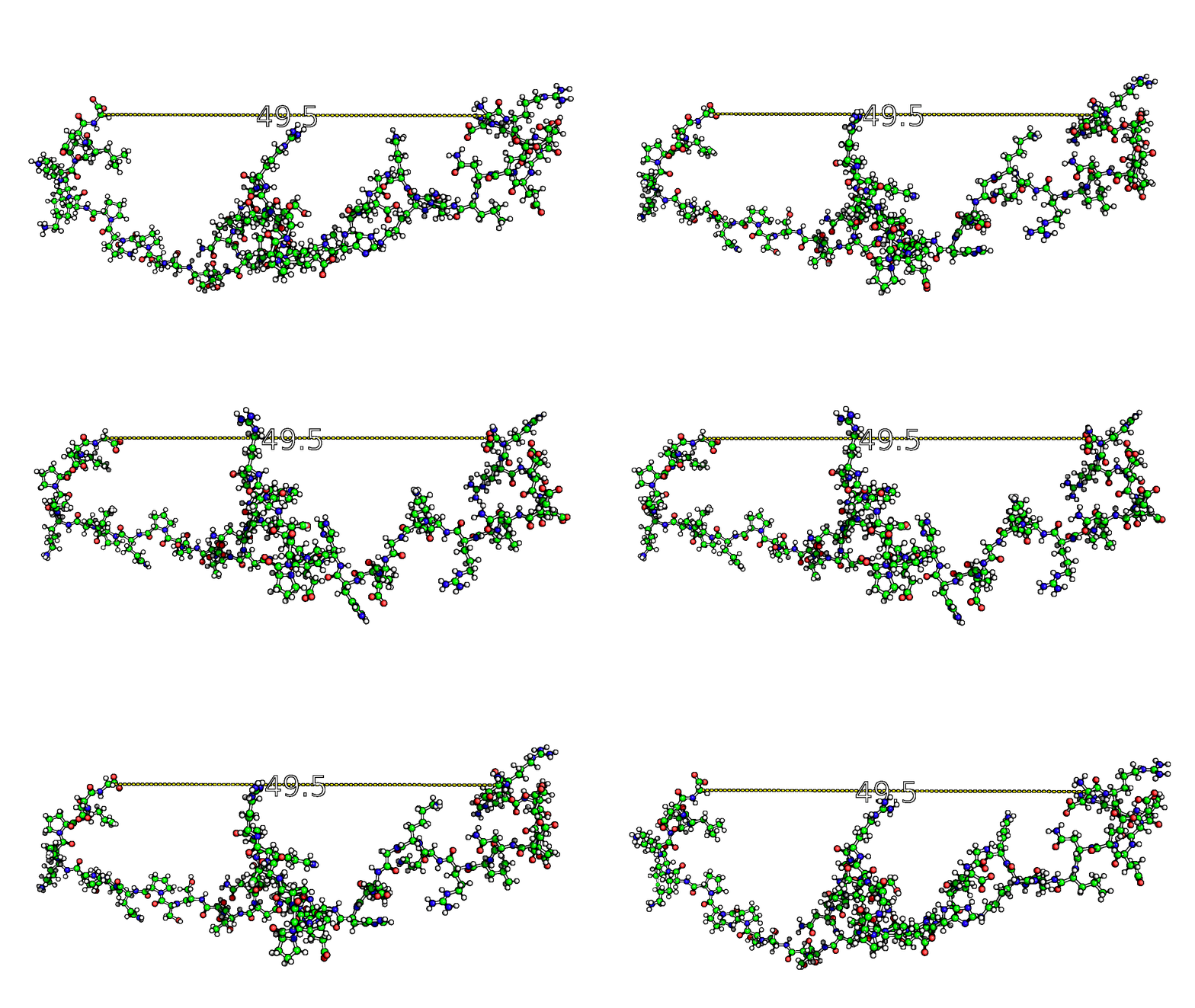
- Tested $\mathbf{End\text{-}to\text{-}End\ Distance\ (EE_{dist})}$ values ranged from the fully contracted ($\mathbf{0.5\ \text{nm}}$) to the expanded state ($\mathbf{7.0\ \text{nm}}$). Crucially, simulations included the distances observed in experimental structures:
- 3.0 nm (Reflecting the p53 monomeric form based on EM/SAXS rigid body model predictions).
- 5.0 nm (Reflecting the p53 tetrameric, DNA-bound form based on EM/SAXS predictions).
Key Findings on Conformational Dynamics and Interactions
1. Improved Agreement with Experimental Data
Restraining the $EE_{dist}$ to experimentally relevant values dramatically improved the model’s accuracy:
- The trajectory simulated with $\mathbf{EE_{dist} = 3.0\ \text{nm}}$ showed the strongest preference and agreement with experimental Nuclear Magnetic Resonance (NMR) chemical shifts for the backbone carbons ($C_{\alpha}$ and $C’$).
- For the backbone carbons, this restraint improved the Root-Mean-Square Error (RMSE) by $\mathbf{0.22\ \text{nm}}$ for $C_\alpha$ atoms compared to the unrestrained trajectory.
- This success highlights the importance of restraining the $EE_{dist}$ when studying IDRs that act as linkers between globular domains.
2. Secondary Structure Transition with Expansion
The conformational landscape is greatly altered by restricting the end terminals. Evaluating the secondary structure shows a clear $\mathbf{structural\ shift\ upon\ expansion}$:
- Contraction ($\mathbf{0.5\ \text{nm}}$): Favors more globular-related secondary structures like $\mathbf{3{10}/\alpha}$-helices and $\mathbf{\beta}$-structures, which decrease as the $EE{dist}$ increases.
- Expansion ($\mathbf{>3.0\ \text{nm}}$): Favors PPII helices and Random Coils, which increase with $EE_{dist}$. PPII helices, in particular, increase from $\sim 10\%$ at $0.5\ \text{nm}$ to $\sim 21\%$ at $7.0\ \text{nm}$. This suggests that stabilization at extended states depends more on structures typically associated with disordered regions.
3. Shift in Intramolecular Contacts
The location of intramolecular interactions is sensitive to the $EE_{dist}:
- Contracted States ($\mathbf{0.5\ \text{nm}}$): Interactions are generally between distant residues.
- Expanded States ($\mathbf{>5.0\ \text{nm}}$): Interactions show a preference for more local, terminal contacts.
This investigation provides a better understanding of the PTL’s conformational dynamics and demonstrates that the physiological restraints imposed by its flanking globular domains are essential to accurately model its local interactions and conformational ensemble.
IV. REG: Regulatory Domain (Residues 350–393)
The C-terminal REG contains multiple phosphorylation sites that stabilize the active p53 tetramer.
- Effect of Phosphorylation Level:
- Single phosphorylation ($\mathbf{REG_{SP}}$ at S392): This state closely resembles the tetrameric (active) form. It introduced stabilizing salt bridges between distant residues, such as $R_{363}$ and $K_{381}$.
- Multisite phosphorylation ($\mathbf{REG_{FP}}$): This state significantly decreased the domain size, resembling the monomeric (dysfunctional) form. The long-range salt bridges were discouraged, replaced by less dynamic local interactions.
- Structural Disruption: Multisite phosphorylation significantly destabilized traditional secondary structures, increasing random coil formation and hindering beneficial conformational states observed in the single phosphorylated REG simulation.
Project Collaborators

Marie Skepö's expertise in computational chemistry was critical to this project, given her specialization in modeling intrinsically disordered proteins (IDPs) through MD simulations. She overseed the extensive MD simulations of the four p53 IDRs (TAD1, PRD, PTL, and REG), ensuring the use of accurate force fields, and developing the computational strategies necessary to interpret their complex conformational landscapes, including the effects of post-translational modifications (PTMs) and physical constraints.
