p53 conference poster · Supplementary analysis
How sequence context changes AlphaFold3 predictions of p53
A four-context AlphaFold3 pilot comparing the same p53 residues 1–28 against structural and NMR observables.
Exploratory supplementary pilot · Benchmark manuscript resubmitted
Short answer
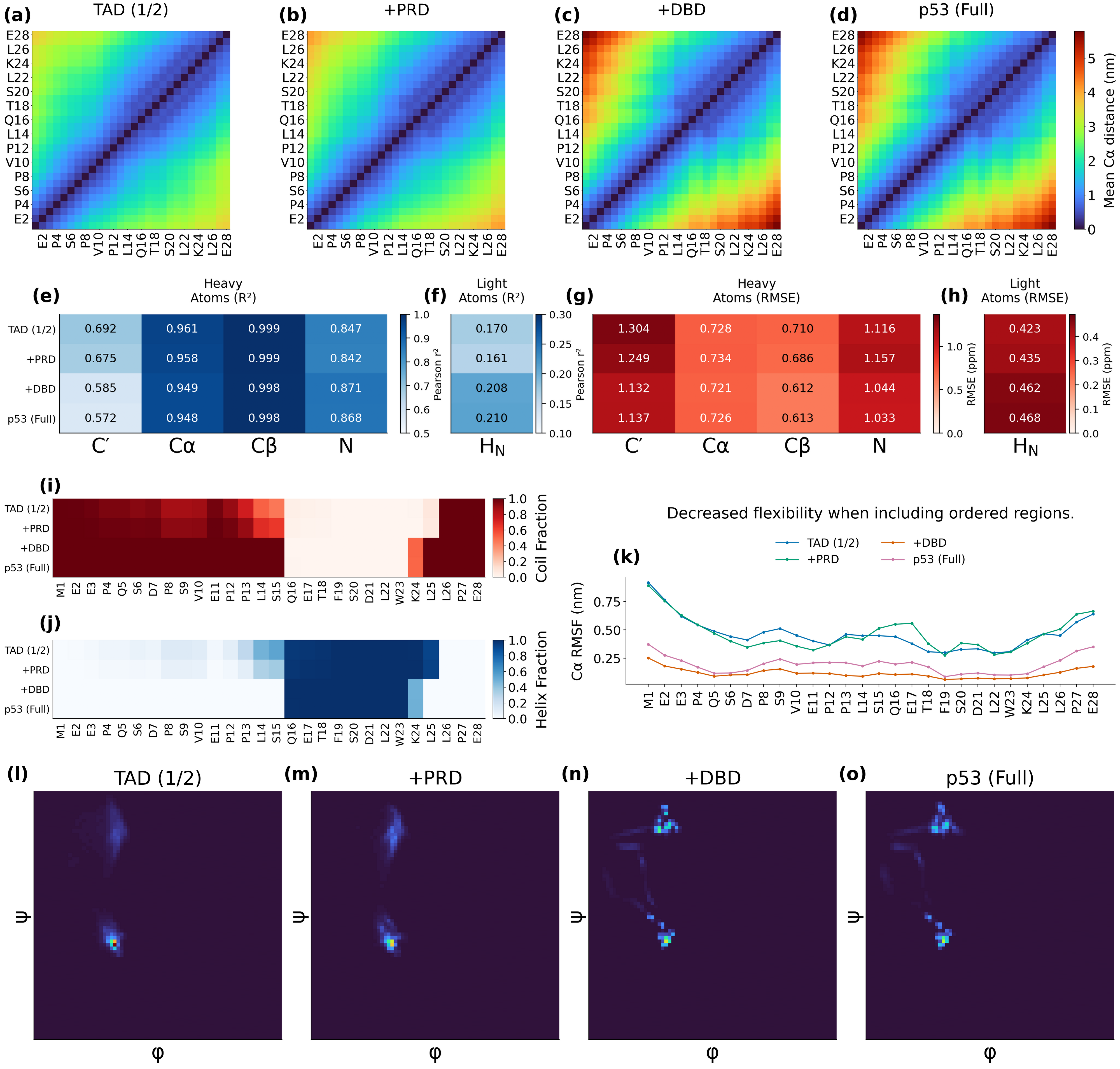
Including more p53 sequence context changed AlphaFold3 predictions for the same N-terminal residues, but did not uniformly improve or worsen agreement with experimental chemical shifts. Larger contexts produced a greater mean distance between residues 1 and 28; changes in chemical-shift agreement depended on the nucleus and metric.
Why context matters
Generative structure models (GSMs) predict molecular conformations, and neighboring sequence or folded domains may influence their outputs. This pilot tests AlphaFold3 only, rather than all six GSMs in the benchmark.
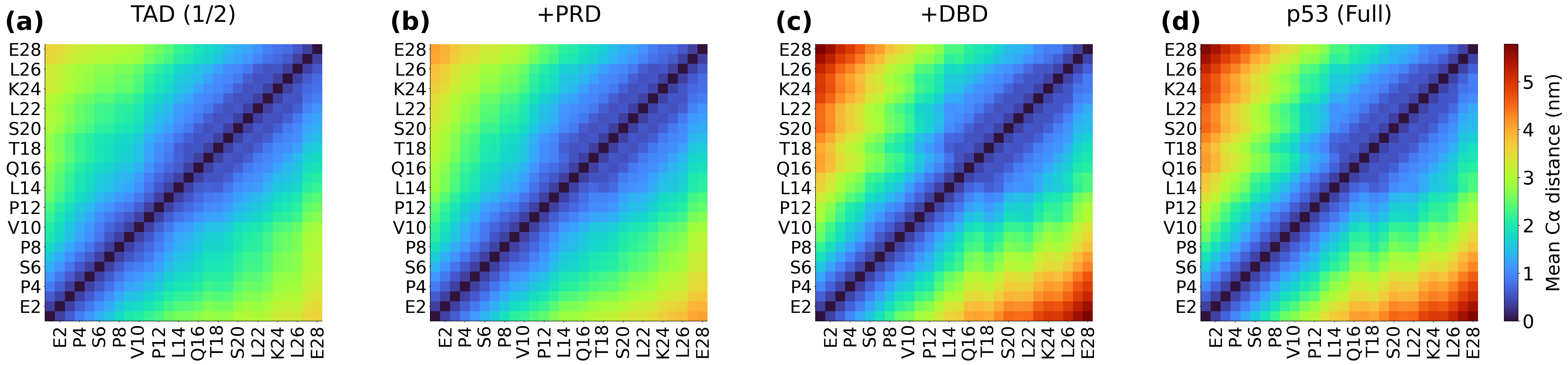
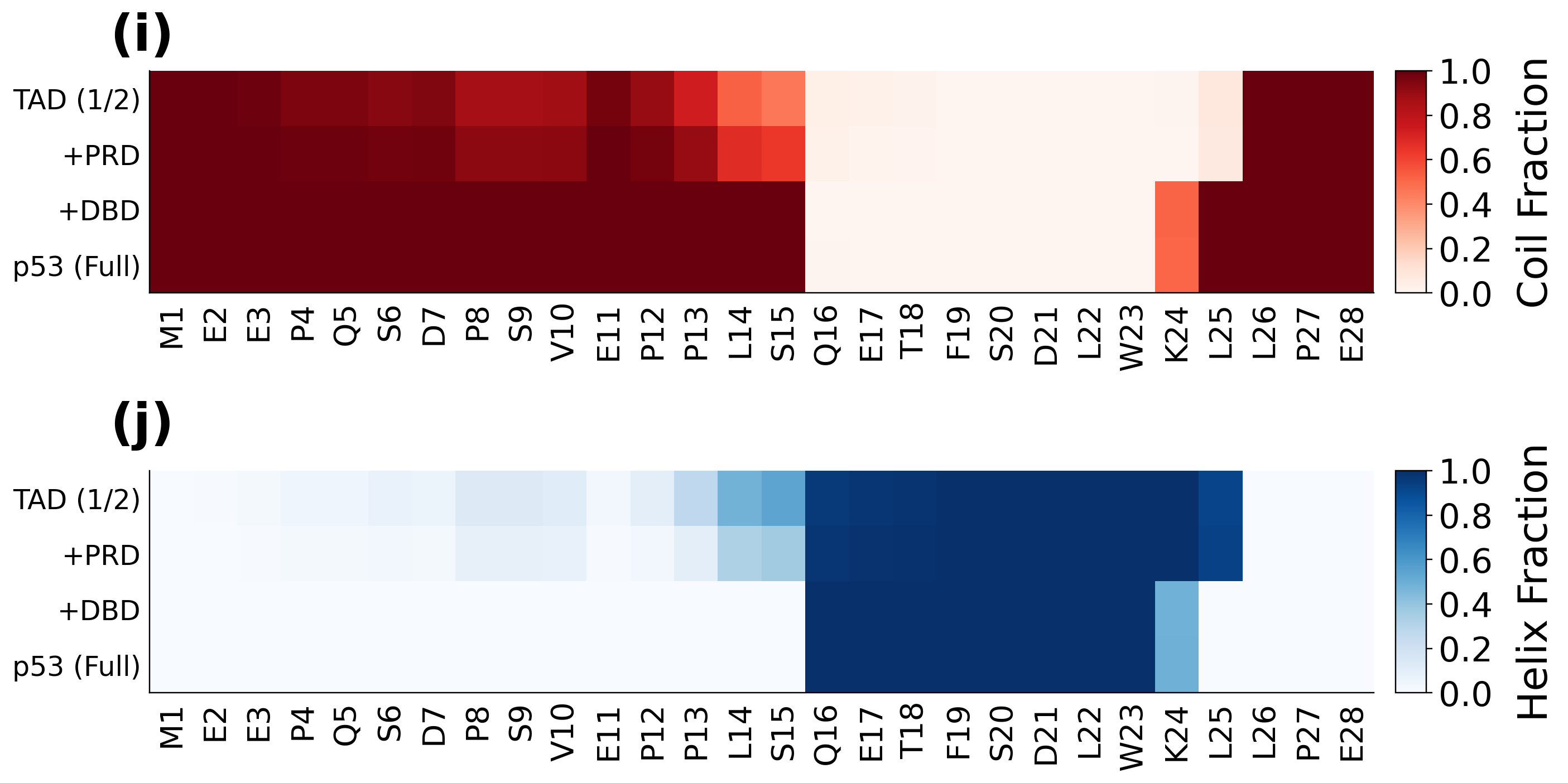
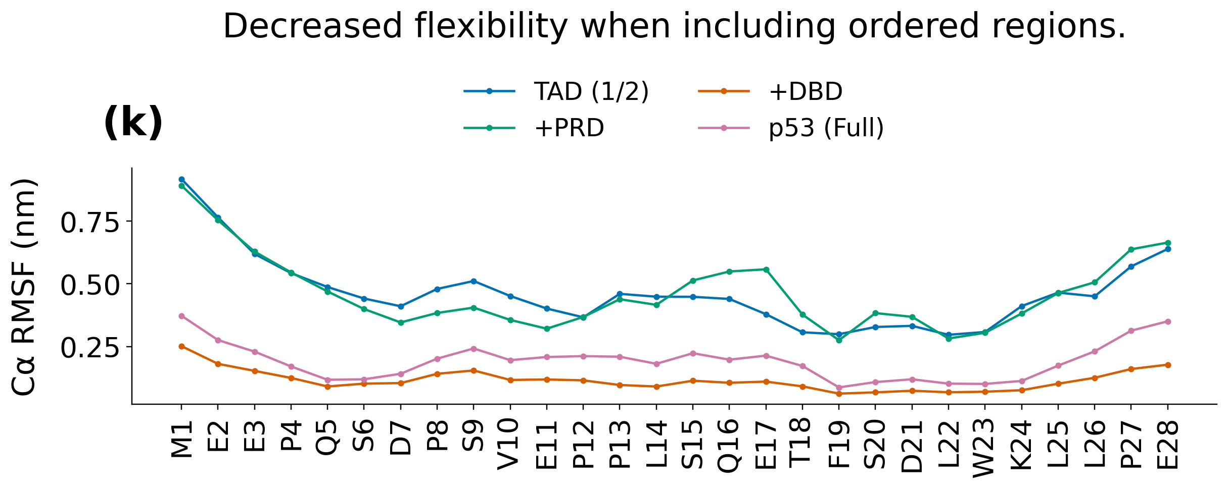
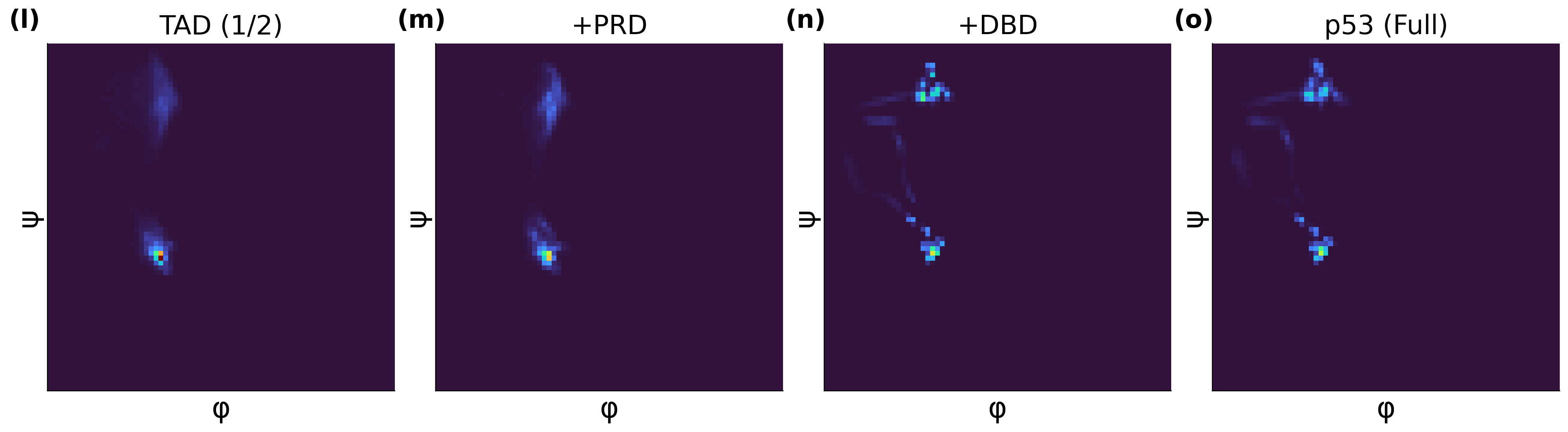
We generated ensembles in four sequence contexts: the transactivation domains (TAD1/2); TAD1/2 plus the proline-rich domain (PRD); those regions plus the DNA-binding domain (DBD); and full-length p53. Every structural and chemical-shift comparison uses the same stored residues 1–28. The smallest context is TAD1/2, not an isolated TAD1 or isolated-1–28 baseline. The width of a stored array does not establish the biological length of its original prediction construct.
Quantitative comparison
Nuclear magnetic resonance (NMR) chemical shifts probe local environments. Root-mean-square error (RMSE) measures absolute prediction error; chemical-shift errors below are in parts per million (ppm). The distance is the ensemble-mean Cα distance from residue 1 to residue 28, in nanometers (nm). Conformation counts are not counts of independent dynamical observations.
| Prediction context | Conformations | Distance 1–28 (nm) | C′ RMSE (ppm) | Cα RMSE (ppm) | Cβ RMSE (ppm) | N RMSE (ppm) | HN RMSE (ppm) |
|---|---|---|---|---|---|---|---|
| TAD1/2 | 4,950 | 3.60 | 1.304 | 0.728 | 0.710 | 1.116 | 0.423 |
| TAD1/2 + PRD | 4,995 | 4.08 | 1.249 | 0.734 | 0.686 | 1.157 | 0.435 |
| TAD1/2 + PRD + DBD | 4,620 | 5.79 | 1.132 | 0.721 | 0.612 | 1.044 | 0.462 |
| Full-length p53 | 4,419 | 5.74 | 1.137 | 0.726 | 0.613 | 1.033 | 0.468 |
Distance maps
Chemical-shift agreement

Secondary-structure fractions
Positional variability
Backbone dihedral distributions
Interpretation
- Mean distance 1–28 increases from 3.60 nm in TAD1/2 to 5.74–5.79 nm in the larger contexts.
- Between TAD1/2 and full-length p53, C′ RMSE improves from 1.304 to 1.137 ppm, whereas HN RMSE worsens from 0.423 to 0.468 ppm. Cα RMSE changes little.
- C′ squared Pearson correlation falls from 0.692 to 0.572, even as its RMSE improves. Association and absolute agreement are different tests. The metrics CSV also reports predictive R², which is a different quantity and may be negative.
- Mean DSSP helix fraction over residues 1–28 falls from approximately 42% to 30% in the larger contexts. Reduced positional variability does not imply greater helicity.
Methods and limitations
The experimental reference is BMRB 17760: wild-type human p53 residues 1–93, pH 7.4, 293 K, ionic strength 175 mM. We compare it with analyzed residues 1–28, so this is not construct-matched validation. Both supplementary pilots use this entry; the primary benchmark uses BMRB 51984.
There are 124 common assignments per context: 26 C′, 27 Cα, 27 Cβ, 22 N, and 22 HN. Experimental H maps to predicted HN; this reference has no Hα comparison. Absolute predicted shifts are averaged with equal conformer weights, without a fitted offset or random-coil subtraction.
RMSF depends on alignment. These descriptive summaries establish neither a causal mechanism, statistical significance, equilibrium populations, nor complete equilibrium sampling. They do not validate binding mechanisms or every observable, and the context trends cannot be generalized to other GSMs.
For a new disordered target, start with a small context pilot. Compare the same residue window and relevant experimental observables: a larger prediction context is not automatically better.
Full overview figure
Downloads and related study
The study dataset accompanies the p53 generative structure model benchmark. This does not imply that every additional website pilot asset or script is deposited there. Downloadable originals on this page are preserved unchanged; asset checksums are available for verification.
Related analysis: Explore the companion study.
Study authors: Michael Bakker, Oskar Svensson, and Marie Skepö.
Contact: Michael Bakker. Back to research.